
Rehabilitation of Ankle and Foot Injuries in Athletes

Rehabilitation of Ankle and Foot Injuries in Athletes
Abstract
Foot and ankle injuries are extremely common among athletes and other physically active individuals. Rehabilitation programs that emphasize the use of therapeutic exercise to restore joint range of motion, muscle strength, neuromuscular coordination, and gait mechanics have been shown to have clinical success for patients suffering various foot and ankle pathologies. Rehabilitation programs are discussed for ankle sprains, plantar fasciitis, Achilles tendonitis, and turf toe.
Introduction
The foot and ankle are among the most common sites for both acute and chronic injuries in athletes and other physically active individuals. 1 Although seldom life-threatening, they often have detrimental effects on sport activity and participation. When an injury to the foot or ankle occurs athletes are limited in their abilities to run, jump, kick, and change directions. Thus, the treatment and rehabilitation of these injuries are crucial in returning athletes to full participation at full functioning. When managing injuries for the foot and ankle, all of the typical clinical considerations must be thought of (type of injury, severity, healing time, type and level of activity, etc), but it is also important to consider other factors such as foot type, biomechanics, footwear worn during activity, and external supports such as bracing or taping. The foot is the base of the lower quarter kinetic chain, thus if rehabilitation and treatment is not managed properly, an injury to the foot or ankle can ultimately cause secondary injuries elsewhere up the chain.
Biomechanics of normal walking
For all sports medicine specialists, evaluation of gait is important for the rehabilitation of lower extremity injuries. Understanding the normal gait pattern will enable a clinician to identify and correct improper compensations after injury. The identification of gait abnormalities should play a key component in deciding to refer a patient for supervised rehabilitation. The movement of the lower extremity during normal walking and running can be divided into two phases, the stance phase and the swing phase.
The stance or support phase, starts with initial contact at heel strike and ends at toe-off. This phase has two important functions. First, at heel strike, the foot acts like a shock absorber to the impact forces and then the foot adapts to the surface. Secondly, at toe-off the foot functions as a rigid level to transmit the force from the foot to the surface. At initial contact, the subtalar joint is supinated and there is an external rotation of the tibia. As the foot loads, the subtalar joint moves into a pronated position until the forefoot is in contact with the ground. The change in subtalar motion occurs between initial heel strike and 20 percent into the support phase of running. As pronation occurs at the subtalar joint the tibia will rotate internally. Transverse plane rotation occurs at the knee joint because of this tibial rotation. Pronation of the foot unlocks the midtarsal joint and allows the foot to assist in shock absorption and to adapt to the uneven surfaces. It is important during initial impract to reduce the ground reaction forces and to distribute the load evenly on many different anatomical structures throughout the foot and leg. Pronation is normal and allows for this distribution of forces on as many structures as possible to avoid excessive loading on just a few structures. The subtalar joint remains in a pronated position until 55 to 85 percent of the support phase with maximam pronation is concurrent with the body’s center of gravity passing over the base of support. From 70 to 90 percent of the support phase, the foot begins to resupinate and will approach the neutral subtalar position. In supination the midtarsal joints are locked and the foot becomes stable and rigid to prepare for push-off. This rigid position allows the foot to exert a greater amount of force from the lower extremity to the surface. The swing phase begins immediately after toe-off and ends as just prior to heel-strike. During the swing phase the leg is moved from behind the body to a position in front of the body. 2
Lateral ankle sprain
Lateral ankle sprains are common acute injuries suffered by athletes. 1, 3 The most common mechanism for a lateral ankle sprain is excessive inversion and plantar flexion of the reafoot on the tibia. The injured ligaments are located on the lateral aspect of the ankle and include the anterior talofibular, the posterior talofibular, and the calcaneofibular. 4
With lateral ankle sprains, the severity of the ligament damage will determine the classification and course of treatment. In a grade 1 sprain, there is stretching of the ligaments with little or no joint instability. Pain and swelling for a grade 1 sprain are often mild and seldom debilitating. After initial management for pain and swelling of the grade 1 sprain, rehabilitation can often be started immediately. Time loss from physical activity for a grade 1 sprain is typically less than one week. Grade 2 sprains occur with some tearing of ligamentous fibers and moderate instability of the joint. Pain and swelling are moderate to severe and often immobilization is required for several days. With a grade 3 sprain, there is total rupture of the ligament with gross instability of the joint. Pain and swelling is so debilitating that weight bearing is impossible for up to several weeks. 5
Rehabilitation Expectations
With lateral ankle sprains regaining full range of motion, strength, and neuromuscular coordination are paramount during rehabilitation. Isometrics and open-chain range of motion can be completed by those patients who are non-weight bearing. Range of motion should focus on dorsiflexion and plantar flexion and be performed passively and actively as tolerated. During early rehabilitation, towel stretches, and wobble board range of motion should be introduced as tolerated. Stationary biking can aid dorsiflexion and plantar flexion motion in a controlled environment while also providing a cardiovascular workout for the athlete. Clinicians can also incorporate joint mobilizations to aid in dorsiflexion range of motion. 6 Hydrotherapy is an excellent means to work on range of motion while also gaining the benefits of hydrostatic pressure.
Once weight bearing is tolerated, middle stage rehabilitation is started. This includes balance and neuromuscular control exercises as well as continued range of motion exercises as tolerated. Balance activities should progress from double-limbed stance to single-limb stance as well as from a firm surface to progressively more unstable surfaces. (see figure 1). Closing the eyes or incorporating perturbations can further challenge patients. Patients can be asked to throw and catch weighted balls, perform single leg squats, and perform single limb balance and reaching exercises. 7 Regaining and maintaining range of motion should be continued. Wobble board training and slant board stretches are also important to focus on heel cord stretching.

Increased strengthening exercises should be started once swelling and pain is controlled. Initially, dorsiflexion and plantar flexion strength should be focused on. Weight bearing calf raises and squats are examples excellent beginning exercises. As the ligaments heal, inversion and eversion strengthening should be added as tolerated. Resistance bands and ankle weights are a good means to gain strength in all planes of motion. (see figure 2) Clinicians can integrate diagonal exercises (ie, combined plantar flexion/inversion and dorsiflexion/eversion) to isolate motions at the talocrural joint.
During this time, it is paramount for clinicians to re-educate athletes on the proper mechanics of walking. Once range of motion and strength is regained, functional activities are included. Functional rehabilitation exercises should begin with simple, uniplaner exercises; walking and jogging in a straight line. Once the athlete can perform these without a pain or a limp, hops, jumps, skips and change of direction can start to be added. Have the athlete perform 10 jumps for distance on the uninvolved limb and challenge him/her to match the distance with the involved limb. Do the same for jumps for height. As cutting is started, begin with wide arching turns and progress to tighter, sharper and faster cuts. Athletes should be challenged to perform lateral movements as well such as shuffling and carioca. As the patient becomes more comfortable and functional, have him/her perform rehab wearing the typical shoe/cleat for the sport and progress to more sport specific activities.
Depending on the severity of the ankle sprain, fear avoidance may cause the athlete to alter play and be at higher risk for reinjury or injury to another location. Also, some sport-specific skills may need to be reconditioned. Participation into sport should start with non-contact drills and progress to contact drills and finally to full scrimmage.
Criteria for full competition
Full return to activity should be a gradual progression in order to stress the ligaments without causing further harm. Full activity should be allowed once the athlete has complete range of motion, 80 to 90 percent of preinjury strength, and a normal gait pattern including the ability to perform sport-specific tasks such as cutting and landing without any compensations due to the injury.. The athlete should be capable, without pain or swelling to complete a full practice.
Clinical Pearls
-
Challenge patients with home-exercises. Have them try to balance on involved limb while brushing teeth, progress to eyes shut while brushing their teeth and balancing
-
Have rehabilitation clinicians perform talocrural and tibiofibular joint mobilizations to increase dorsiflexion
-
Perform exercises with shoes on and off to alter the planter cutaneous feedback
-
Using a 10-20 yard area, have the patient walk on toes back in forth. Repeat walking with toes pointed in, toes pointed out, and on heels (toes in the air)
-
Ask the subject to perform 10 single leg jumps in a row on the involved limb as high as possible in a row, while watching his/her face, if he/she can complete without grimacing safe to start functional rehabilitation.
Patient Education
The leading predisposing factor for an ankle sprain is a history of an ankle sprain 8 and an estimated 30% of all individuals who suffer an initial ankle sprain will develop chronic ankle instability. 4 Thus, patients need to know that inadequate treatment and rehabilitation of an ankle sprain has a great likelihood of leading to future problems.
Often patients, coaches, and parents have the mindset that a lateral ankle sprain is not serious and players can return quickly. In fact, in many cases health care services are not even sought by individuals suffering an ankle sprain. 9 It is critical for all stakeholders to understand the high frequency of residual symptoms and recurrent sprains. 10 The importance of allowing the ligaments to heal, regaining full range of motion, strength, and balance prior to returning to activity must be emphasized to patients. If, while doing rehabilitation swelling returns, patients must know that they did too much.
Prophylactic support
Prophylactic support is often used after an ankle sprain to provide mechanical stability. Depending upon individual preference and budget, athletes can use tape or a variety of braces (lace-up, stirrup, or elastic type of configuration. Both taping and bracing have been found to reduce the risk of recurrent ankle sprains in athletes. 11 Advantages of a braces include easy of application and cost effectiveness. Braces also provide the athlete with proprioceptive stimulation, which implies an improve proprioception and sensory feedback. 12 Taping, one the other hand can be custom designed for the specific athlete, sport, and instabilities.
Other ankle sprains
Although less common, medial and syndesmodic ankle sprains often result in more severe injuries causing longer time to heal and rehabilitate. Medial ankle sprains occur with a mechanism of excessive eversion and dorsiflexion, causing the deltoid ligament to be injured. Patients with medial ankle sprains will often present with swelling and discoloration on the medial aspect of the ankle and unwillingness to bear weight.
Syndesmodic sprains occur with disruption of the interosseous (or syndesmodic) ligament that stabilizing the inferior tibiofibular joint. Injury to this ligament occurs with excessive external rotational or forced dorsiflexion. Syndesmotic sprains may occur in isolation or in combination with medial or lateral ankle sprains. Due to limited blood supply and the difficulty in allowing the injured ligament to heal unless the ankle is immobilized, injuries to the syndesmodic ligaments often take months to heal. 13 Patients with syndesmodic sprains often present with a lack of swelling, but will be extremely tender over anterior aspect of the distal tibiofibular joint.
Rehabilitation expectations
Initial treatment for both medial and syndesmodic sprains is often immobilization and crutches. During this time, swelling and pain management are the primary concerns. The length of time of immobilization will vary among patients and will depend on the severity of the sprain. While immobilized, patients can work on controlled open-chain range of motion, focusing on dorsiflexion and plantar flexion. During this time, inversion and eversion should be held to a minimum. During early rehabilitation, nothing should increase pain or swelling to the area.
Once weight-bearing is tolerated, crutches should be used at a minimum. Gait training may be needed to ensure the patient is not compensating in any way, which may cause secondary injury. At this point, rehabilitation will follow the progression as stated above in the lateral ankle section. Rehabilitation concerns include; pain and swelling, range of motion, strength, balance and neuromuscular control, and functional exercises.
Criteria for full competition
Full return to activity should be a gradual progression in order to stress the ligaments without causing further harm. Full activity should be allowed once the athlete has complete range of motion, 80 to 90 percent of preinjury strength, and the ability to perform gait activities (including running and changing direction) without difficulty.. The athlete should be capable, without pain or swelling to complete a full practice.
Patient education
With medial and syndesmodic sprains, patience is the most important thing for the patient to learn. The healing of the medial and syndesmodic ligaments take time, sometimes up to severalmonths to fully heal. The difference in the expectations of these injuries compared to lateral ankle sprains must be emphasized to all stakeholders so that realistic expectations for return to play can be understood.
Plantar Fasciitis
Plantar fasciitis is the catchall term that is commonly used to describe pain on the plantar aspect of the proximal arch and heel. The plantar fascia is an aporneurosis that runs the length of the sole of the foot and is a broad dense band of connective tissue. It is attached proximally to the medial surface of the calcaneus and fans out distally, attaching to the metatarsophalangeal articulations and merges into the capsular ligaments. The plantar aponeurosis assists in maintaining the stability of the foot and secures or braces the longitudinal arch.
Plantar fasciitis is caused by a straining of the fascia near its origin. The plantar fascia is under tension with toe extension and depression of the longitudinal arch. During normal standing (weight bearing principally on the heel), the fascia is under minimal stress, however, when the weight is shifted to the balls of the feet (running) the fascia is put under stress and strain. Often planar fasciitis is a result of chronic running with poor technique, poor footwear, or because of lordosis, a condition in which the increased forward tilt of the pelvis produces an unfavorable angle of foot-strike when there is considerable force exerted on the ball of the foot. 14
Patients more prone to plantar fasciitis include: those with a pes cavus foot; excessive pronation; overweight; walking , running or standing for long periods of time, especially on hard surfaces; old, worn shoes (insufficient arch support); and tight Achilles tendon. 14 The patient will present with pain in the anterior medial heel, usually at the attachment of the plantar fascia to the calcaneus. The pain is particularly noticeable during the first couple of steps in the morning or after sitting for a long time. Often the pain will lessen as the patient moves more, however the pain will increase if the athlete is on his/her feet excessively or on his/her toes often. Upon inspection, the plantar fascia may or may not be swollen with crepitus. The patient’s pain will increase with forefoot and toe dorsiflexion.
Rehabilitation expectations
Depending on patient compliance, plantar fasciitis can be a very treatable minor injury with symptoms lasting days. However, without proper treatment and patient compliance, plantar fasciitis can linger for months or even years.
Initial treatment of plantar fasciitis starts with pain control. Rest is extremely important at this time, patients should not being performing any unnecessary weight bearing. Patients should also be wearing comfortable supportive shoes when walking is necessary. Adding a heel cup or custom foot orthosis to a patient’s shoe may relieve some of the pain at the plantar fascia insertion. 15 During this time, regaining full dorsiflexion range of motion of the foot as well as of the big toe is vital. Towel stretches, slant board stretches, and joint mobilizations administered by a rehabilitation clinician will aid in the return of dorsiflexion range of motion.
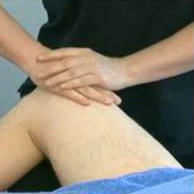
After pain is reduced strengthening exercises can be incorporated into rehabilitatoin. The focus should be in strengthening some of the smaller extrinsic and intrinsic muscles of the foot. Towel crutches, big toe-little toes raises, short foot exercises are good examples of strengthening exercises ( Fig 3). Throughout the treatment and rehabilitation process, soft tissue work such as cross-friction massage may aid in the alleviation of symptoms.

Criteria for full competition
Although athletes can often continue to participate fully while suffering from plantar fasciitis, it should be understood that the longer activity is continued, the longer the symptoms will linger. For best recovery of this injury, extra activity should not be started until the athlete is able to walk a full day without any pain. Once a daily activities are tolerated, activity can slowly be increased until full participation. Throughout the rehabilitation and participation progression stretching should occur often throughout the day.
Clinical Pearls
-
While sitting, roll on a ball (tennis ball, golf ball, etc) underneath the medial longitudinal to stretch the plantar fascia
-
Fill a paper cup with water and freeze it, roll on the frozen cup to get the benefits of cold while also stretching the plantar fascia
-
Before getting out of bed in the morning, but on shoes with good arch supports to provide the plantar fascia support upon weight-bearing
-
Sleep with feet off the end of the bed to allow some dorsiflexion while sleeping
-
Wear a night splint that will keep foot in a dorsiflexed or neutral position
-
Stretching often throughout the day for a short period of time is more beneficial then stretching once a day for a long period of time
-
Do not weight high-heels or other shoes with no support (sandals) during the day
Patient education
Plantar fascia tends to be a cyclical injury. Athletes will repetitively suffer from this injury because after the initial injury, the cause of the injury is not treated, only the symptoms. Patients with plantar fasciitis need to have their gait biomechanics thoroughly evaluated and, if necessary, be fitted for custom orthotics. 15
Achilles Tendonitis
Achilles tendonitis is an inflammatory condition that involves the Achilles tendon and/or its tendon sheath. Achilles tendonitis is the most common overuse injuries reported in distance runners. 16 Although Achilles tendonitis is generally a chronic condition, acute injury may also occur. Typically, the athlete will suffer from gradual pain and stiffness about the Achilles tendon region, 2 to 6 cm proximal to the calcaneal insertion. The pain will increase after running hills, stairs, or an increased amount of sprints (running on toes). Upon evaluation, the gastrocnemius and soleus muscle testing may be normal however, flexibility will be reduced. Having the patient perform toe raises to fatigue will show a deficit compared to the uninvolved limb. Inspection of the area may feel warm to the touch and pain, tenderness and crepitus may be felt with palpation. The tendon may appear thickened indicating a chronic condition.
Rehabilitation expectations
Healing of Achilles tendonitis is a slow process due to the lack of vascularity to the tendon. Initially, patients will feel comfortable by placing less stress to the area by wearing a heel cup. Resting and activity modification is important during the initially healing stages. The clinician needs to emphasize the importance of allowing the tendon to heal. During this time, cross friction massage can be started to the area to break down adhesions and promote blood flow to the area.
Stretching and strengthening of the gastrocnemious-soleus complex should be incorporated as tolerated by the patient. Towel stretching and slant board stretching should be done throughout the day. As range of motion is restored, the heel cup should be removed to reduce the chances of adaptive shortening of the muscles and tendon. Progressive strengthening including toe raises and resistive tubing should be incorporated at the beginning of rehabilitation. Sets should start low with low reps and gradually increase to low sets high reps for endurance as tolerated by the athlete. As pain and inflammation decreases, machine weights, lunges, and sport specific exercises can be added. Eccentric exercises for the triceps surae often have beneficial results in athletes with Achilles tendonitis. 17
The patient’s foot structure and gait mechanics should be evaluated for possible orthotic benefits. Often Achilles tendonitis is a result of overpronation, an abnormality that can be addressed with foot orthoses. 18 Once range of motion, strength and endurance has returned, athletes should slowly progress into walking and jogging program. Workouts should be done on a flat surface when possible. The walking and jogging program should start out with slow mini-bursts of speed. The program is to increase the amount of stress the Achilles tendon can tolerate; it is not to improve overall endurance. As tolerated by the patient, running and sprinting can be increased.
Criteria for full competition
Athletes should be allowed to compete when full range of motion and strength has returned. The athlete should have regained endurance in the involved limb and be capable of completing a full practice without pain. Depending on the sport, some athletes may be able to compete while suffering from Achilles tendonitis. However, patients should be educated in the fact that the condition will not go away without proper rest and treatment.
Patient Education
Patients need to be educated with the risks of Achilles tendonitis, specifically hill running, lack of proper shoes, lack of rest, and flexibility. Hill workouts increase the stress and strain to the gastrocnemius-soleus complex and Achilles tendon. Hill workouts should be done at a maximum once a week to allow the body time to heal. Similar to any chronic injury to the feet, shoes must be evaluated. Athletes need to learn and understand their foot type and the proper shoes for their foot type. Also, shoes should be replaced every 500 miles are a maximum 2 years. Running on old worn shoes will alter biomechanics and cause stress and strain to the body. Finally, the lack of flexibility is often the main culprit in Achilles tendonitis. The importance of stretching and stretching often should be emphasized.
Prophylactic support
Initially, heel cups will reduce the tension and stress placed on the Achilles tendon. As flexibility is regained, the heel cup should be gradually reduced to reduce the chances of an adaptive shortening of the tendon. Athletes may find comfort in a special tape job that will reduce the stress placed on the Achilles tendon as well. The patient’s foot type and gait mechanics should be evaluated for possible use of custom orthotics. Achilles tendonitis can often be attributed to over pronation during gait. A custom orthotic will be able to adjust the athlete’s gait to reduce this abnormality.
Turf Toe
Tuft toe is a hyperextension injury of the great toe, causing a sprain to the metatarsophalangeal joint and damage to the joint capsule. Turf toe can be either an acute or a chronic condition. An acute turf toe often occurs when the athlete’s shoe sticks into the ground while he/she is trying to stop quickly. The shoe sticks as the individual’s body weight shifts forward, causing the big toe to jam into the shoe and ground. The chronic condition occurs from frequent running or jumping in shoes that allow excessive great toe motion. This mechanism of injury may occur on natural or synthetic surfaces. 19
Athletes with turf toe will present with pain at the 1st metatarsophalangeal joint. Swelling and stiffness may be present, however pain, especially with great toe extension is the primary symptom. Rehabilitation of turf toe typically requires several weeks. If left untreated, turf toe can lead to permanent decrease in range of motion and osteoarthritis arthritis. 19
Rehabilitation concerns
Patients suffering from turf toe respond best with rest and an adjustment made to their shoes. Pain management should be of primary concern to the clinician. Once pain and swelling have been reduced, the athlete should start performing toe extension and flexion exercises such as toe crunches and short foot exercises. Joint mobilizations should be added to the treatment protocol to aid in pain and increase range of motion. Once pain and swelling is reduced, the athlete may begin to progress into athletic activities. Protecting the great toe with a stiff forefoot insert or a great toe taping may increase athlete comfort.
Criteria for full competition
Athletes are able to return to full competition when any pain and swelling has resolved. Often athletes with turf toe are capable to continue practicing and participating while suffering from this injury with the toe being taped and possible inserts into shoes.
Pearls of wisdom
-
Have patients wear stiff insoled shoes to prevent excessive motion
-
Great toe joint mobilizations can be incorporated to reduce pain and increase motion
Patient education
Patients should be aware that if left untreated, turf toe may cause permanent decreased range of motion in the great toe and bone spurs may develop. Although athletes can often play with turf toe, rest and pain management is the most beneficial for athletes. Without prevention of excessive extension of the great toe, symptoms of turf toe may disappear with rest just to return once the athlete returns to activity.
Prophylactic support
Athletes with turf toe may benefit from adding a steel or other stiff material insert into the forefoot of the shoes to reduce extension. Taping of the great toe to prevent dorsiflexion may also be done.
Conclusions
Nearly all lower extremity injuries in athletes will benefit from rehabilitation programs that include therapeutic exercise. Restoring joint range of motion, muscle strength, and neuromuscular coordination should be emphasized as should normal gait mechanics. A graduated return to physical activity that includes sports-specific exercises is recommended with the primary goals being to allow a safe return to sport while minimizing the risk of recurrent injuries.
Footnotes
Publisher's Disclaimer: This is a PDF file of an unedited manuscript that has been accepted for publication. As a service to our customers we are providing this early version of the manuscript. The manuscript will undergo copyediting, typesetting, and review of the resulting proof before it is published in its final citable form. Please note that during the production process errors may be discovered which could affect the content, and all legal disclaimers that apply to the journal pertain.

